Advances Across Ophthalmology: A Practical Guide to Geographic Atrophy, Ocular Surface Disease, Glaucoma, and Refractive IOL Surgery
Course is eligible for 1.5 AMA PRA Category 1 Credits™.
Log in to fulfill the requirements for free in our secure environment, the Learning Center.

Emerging Interventions for Geographic Atrophy
Advancing detection, treatment, and patient-centered care.
Faculty:
- Lejla Vajzovic, MD
- Sophie J. Bakri, MD
- Rishi P. Singh, MD
The Clinical and Functional Burden of Geographic Atrophy
By Lejla Vajzovic, MD
Geographic atrophy (GA) is a major clinical challenge in retinal practice. As the advanced form of dry age-related macular degeneration (AMD), GA leads to irreversible and progressive loss of photoreceptors and retinal pigment epithelium (RPE), leading to functional vision loss that significantly impairs patient quality of life. For many, the disease threatens core aspects of independence. Patients experience broad functional impairment, including difficulty reading, reduced driving ability, challenges with daily activities, and reliance on visual aids.1
For many patients, the impact on driving is particularly significant. Among licensed drivers with GA, 50% and 88% reported lacking confidence driving during the day and at night, respectively. Most (82%) reported worsening vision over the past year compared with only 25% of controls (OR 13.55; P < .001).2 Loss of driving independence compounds isolation, limits access to appointments, and erodes the self-sufficiency that defines daily living for many older adults.
GA is also a heterogeneous disease. Progression rates vary substantially across patients, making it difficult to predict clinical course and determine the optimal timing of intervention. The variability highlights the need for consistent monitoring and systematic identification of patients at highest risk for rapid disease progression.
Several clinical parameters help guide risk stratification in practice. Lesion size at baseline is one of the most reliable predictors. Larger areas of GA at initial presentation correlate with accelerated future growth.3–5 Lesion pattern on fundus autofluorescence (FAF) is equally informative. High-risk FAF patterns, including banded and diffuse hyperautofluorescence at lesion borders, are associated with significantly faster growth rates (diffuse: 3.78 mm²/year; banded: 2.52 mm²/year) compared with focal or no hyperautofluorescence patterns (0.36 and 0.02 mm²/year, respectively).3,6
Multifocality is another key prognostic variable. Patients with multiple discrete GA lesions tend to progress more rapidly than those with unifocal disease.7 Extrafoveal and perifoveal lesion location, while often associated with preserved central acuity in the short term, carries its own prognostic concern because they typically expand faster than subfoveal disease, ultimately threatening the fovea.8 The status of the fellow eye also provides important context: When significant progression has occurred in a contralateral eye, symmetric behavior in a treated eye should be anticipated.4 Finally, tracking historical progression rates from prior imaging enables a more individualized forecast of where each patient falls on the progression continuum.5
On OCT, reticular pseudodrusen (RPD), characterized by irregularities above the RPE rather than the sub-RPE deposits seen with classic drusen, represent a high-risk biomarker for progression toward advanced disease.9,10 Their presence should prompt closer monitoring and consideration for earlier intervention.
CONCLUSION
Real-world integration of GA management requires both clinical acuity and honest acknowledgment of current barriers. Even with the recent expansion of approved therapies, early intervention remains underutilized. Therapy access and patient acceptance are among the most frequently cited barriers. These realities demand attention not only at the level of individual patient counseling, but in the broader systems and workflows that govern retinal care
Innovations in Earlier Detection
By Sophie J. Bakri, MD, MBA
The case for multimodal imaging in GA is straightforward. No single imaging modality provides a complete picture of disease status, progression risk, and treatment response. Each modality provides complementary information, enabling clinicians to move from descriptive assessment toward more predictive disease management.
FUNDAMENTALS FOR SUCCESS
Currently, OCT is the cornerstone of GA imaging and monitoring in most clinical settings. It enables direct visualization and quantification of structural changes central to GA pathophysiology, including drusen characterization, hyperreflective foci, ellipsoid zone (EZ) loss, and RPE atrophy (Figure 1).

Figure 1. OCT helps detect early structural changes associated with GA
OCT-derived quantitative metrics such as lesion size and the ratio of photoreceptor to RPE loss have emerged as important predictors of progression velocity and treatment response; a higher photoreceptor-to-RPE loss ratio is associated with greater responsiveness to complement inhibitor therapy.11
Incomplete outer retinal atrophy (iRORA) and complete outer retinal atrophy (cRORA) represent distinct structural stages on the OCT continuum whose differentiation carries both diagnostic and prognostic weight. Their classification should be based on careful structural assessment of retinal layers rather than transillumination profiles alone. Importantly, all major OCT platforms now include tools that allow clinicians to track lesion proximity to the fovea and monitor GA size serially. These images may be shown to patients to facilitate understanding of their disease trajectory.
Infrared imaging is a practical workhorse. Its ease of acquisition, compatibility with standard OCT platforms, and ability to clearly delineate GA lesion borders make it useful for serial monitoring and providing a retrospective window into disease history from archived imaging.
FAF adds critical information about lesion margin behavior (Figure 2). Hyperautofluorescence at the borders of a GA lesion signals metabolic stress in adjacent RPE cells and is associated with higher likelihood of near-term lesion expansion.3,6 FAF is particularly valuable in the context of multifocal disease, where it can help identify the full geographic distribution of atrophic areas. The stark hypoautofluorescence of atrophic zones also serves as a vivid and effective patient communication tool.

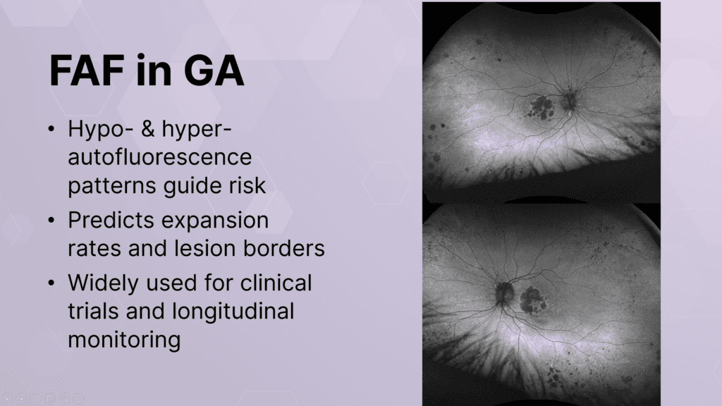
Figure 2. FAF helps predict lesion expansion rates and lesion borders.
Choriocapillaris flow deficits detectable on OCT angiography (OCT-A) represent an early biomarker for disease activity. The modality allows visualization of vascular alterations at the margins of GA lesions with predictive value for expansion rates.12 It can also serve as an efficient screening step to identify subclinical neovascularization before proceeding to fluorescein angiography (FA). This is an important consideration given that patients with GA face substantial risk of conversion to neovascular AMD.
Wide-field imaging complements macular-focused assessments. The imaging of peripheral GA lesions and subclinical changes outside the central field enable a more comprehensive survey of total retinal disease burden.
Emerging evidence suggests that GA may not be strictly a macular condition, and punched-out atrophic lesions can also appear in the retinal periphery, reflecting a broader pathophysiologic milieu that macular imaging alone may miss.13
In clinical practice, the use of both OCT and FAF is common given their complementary structural and metabolic information and their practical accessibility in most retinal practices. Standardizing imaging protocols across visits and offices improves reproducibility and supports more reliable longitudinal comparisons. Further, AI-assisted imaging analysis is transforming the ability to detect and monitor GA. AI-enabled OCT mapping of photoreceptors and RPE supports detection of early disease conversion, quantification of therapeutic response, and monitoring of disease progression over time.11 The photoreceptor-to-RPE loss ratio, calculable from AI-assisted OCT analysis, has been validated as a clinically meaningful predictor and is being integrated into commercial OCT platforms from multiple manufacturers. These tools hold promise for standardizing risk stratification and identifying fast progressors earlier and may have direct implications for treatment timing decisions.
Functional OCT, which integrates microperimetry with structural imaging to monitor retinal sensitivity longitudinally, represents another emerging frontier with potential to capture functional losses that precede visible structural change.
Therapeutic Advances in GA
The case for earlier intervention.
By Rishi P. Singh, MD
The therapeutic landscape for GA is evolving. Long-term data from pivotal clinical trials now inform increasingly nuanced treatment protocols. Two complement inhibitors, pegcetacoplan (targeting C3) and avacincaptad pegol (ACP, targeting C5), have received FDA approval for the treatment of GA.
Pegcetacoplan was evaluated across 48 months of follow-up in the OAKS, DERBY, and GALE open-label extension studies (Figure 3).14 Continuous treatment was found to significantly preserve retinal tissue with greater effectiveness observed in years 3 and 4 than in the initial 2. This finding was consistent with the cumulative, disease-modifying nature of complement inhibition. The early-versus-delayed treatment comparison is particularly instructive: Patients who initiated treatment early preserved 3.16 mm² of retinal tissue at month 48, compared with 1.11 mm² in those whose treatment was delayed.

Figure 3. Results of the OAKS, DERBY, and GALE trials for pegcetacoplan.
To place this in clinical context, this difference approximates more than one disc area of preserved retinal tissue. This result is meaningful in terms of functional vision and the prevention of foveal encroachment. Early-treated patients also demonstrated a 35% risk reduction in vision loss, particularly in critical central vision loci. The safety profile of pegcetacoplan is well-established, with more than 700,000 injections administered in real-world practice.14
The rate of intraocular inflammation (IOI) is approximately 1 in 4,000 injections. To mitigate volume-related IOP spikes, ocular massage before injection is a practical technique. The risk of ischemic optic neuropathy observed in some patients in monthly dosing arms was not observed with every-other-month (EOM) dosing,14 which is currently the preferred regimen for most patients in clinical practice.
Avacincaptad pegol data from GATHER1 and GATHER2 have further established the clinical utility of C5 inhibition (Figure 4). A post-hoc analysis demonstrated a 59% overall risk reduction in the rate of vision loss (defined as ≥15-letter loss in BCVA from baseline at any 2 consecutive visits) compared with sham treatment at 12 months.15 More than 400,000 vials of ACP have been delivered since approval.

Figure 4. Results of the GATHER1 and GATHER2 trials for avacincaptad.
CHALLENGES IN THE CURRENT LANDSCAPE
Several important challenges characterize the current therapeutic landscape. First is the requirement for monthly or bimonthly intravitreal injections. Additional challenges include the potential for intraocular inflammation, volume-related injection side effects including transient IOP elevation, and the fact that current therapies preserve tissue rather than restore vision. Efficacy also appears to vary meaningfully based on patient phenotype, underscoring the importance of individualized treatment planning (see Treatment Efficacy).
Treatment Efficacy
Patient selection and phenotype-based decision making are increasingly central to GA management. For patients with subfoveal GA in one eye and intermediate AMD in the fellow eye, treatment should be considered to preserve vision as long as possible. Vigilant monitoring of the fellow eye is mandatory. Approximately 30% of eyes with central GA develop neovascular AMD within 4 years.16 For fovea-involving GA with retained vision, treatment should be initiated as early as possible to delay further lesion growth. In patients with non-subfoveal multifocal GA lesions, which carry a higher risk of rapid progression given their extrafoveal location, early treatment initiation is warranted to spare the fovea for as long as possible. Elderly age alone should not preclude treatment; phase 3 trial populations had mean ages of 79 and 77 years in the OAKS/DERBY and GATHER2 studies, respectively.16
The timing of intervention is among the most consequential clinical decisions in GA management. A lesion-to-fovea distance of 100 to 200 µm represents a clinically meaningful threshold at which treatment urgency increases substantially. When bilateral disease is present, consideration should be given to treating the better-seeing eye first rather than the worse-seeing eye as is conventional in neovascular AMD management.
A phenotype-based approach supported by photoreceptor-to-RPE loss ratio analysis will increasingly guide both the initiation and monitoring of therapy as these AI-assisted tools become more widely available in commercial OCT platforms.11
Patient-Centered Approaches to Communication and Compliance
Effective treatment of GA is inseparable from effective patient communication.
By Sophie J. Bakri, MD
Unlike neovascular AMD, where patients may experience relatively rapid functional improvement following anti-VEGF therapy, GA treatment offers no visible or subjective benefit in the short term. Rather, the therapeutic goal is deceleration of an inexorable process. This fundamentally different proposition requires a fundamentally different communication strategy.
An analogy to glaucoma management is apt: Compliance is the determinant of long-term outcomes. Achieving compliance, however, depends on patients and families understanding the nature of the disease and the mechanism of benefit. As the long-term data clearly demonstrate,14,15 the cumulative tissue preservation benefit of complement inhibitor therapy increases over time. Early initiation and sustained adherence therefore are critical to achieving optimal outcomes.
BUILDING A SOLID FOUNDATION
Patient education. Informed consent should begin before a treatment decision is made. This requires a clear, jargon-free explanation of AMD, its causes, and its impact on vision. Visual aids, diagrams, and the patient’s own annotated imaging help support patient education.17,18 Patients benefit from understanding the stages of AMD progression and the associated changes in vision they may experience at each stage.17 Educating patients about common symptoms including blurry, distorted vision, difficulty reading, and changes in color perception and encouraging regular self-monitoring through Amsler grid and at-home devices helps patients actively participate in surveillance of their own disease course.
Expectations setting. Setting realistic expectations is equally important. Patients must understand complement inhibitor therapy slows the growth of atrophic lesions but does not restore lost tissue or reverse vision loss already incurred. Framing the treatment goal as vision preservation rather than recovery helps align patient expectations with clinical reality and reduces the disappointment that may otherwise undermine long-term adherence.
Family involvement. Family is a practical and often underutilized resource. Including them in counseling discussions reinforces treatment rationale between visits, assists with appointment logistics, and provides the social support that sustains engagement with a chronic treatment regimen. Documenting the participation also reflects sound medicolegal practice.
Workflow and team-based care. The educational demands of these discussions are substantial and often exceed the capacity of standard clinic appointments. Leveraging the full care team, including optometrists, ophthalmic technicians, nurses, and residents or fellows to deliver educational content and answer follow-up questions can meaningfully expand the practice’s educational capacity.17 Pre-visit educational materials, online modules, and take-home brochures offer additional avenues for preparing patients to engage more productively in clinical conversations.
Patient resources. Low vision rehabilitation and community resources should not be overlooked as essential components of the GA care plan. Magnifiers, contrast-enhancing devices, adaptive lighting, and structured programs through low vision specialists can meaningfully improve daily function and quality of life.17 Encouraging patients to remain socially and physically active and to access available support services addresses the broader functional and psychological burden of the disease that pharmacologic therapy alone cannot fully meet.1
CONCLUSION
As complement inhibitor therapy reshapes the treatment landscape for GA, our ability to communicate its purpose, limitations, and long-term value will be equally decisive in determining patient outcomes. Building a practice culture that prioritizes education, realistic expectation-setting, and sustained engagement supported by the full care team is not ancillary to GA management but rather central to it.
References
2. Patel PJ, et al. Clin Ophthalmol. 2020;14:15.
3. Holz FG, et al. Am J Ophthalmol. 2007;143:463-472.
4. Lindblad AS, et al. Arch Ophthalmol. 2009;127:1168-1174.
5. Sunness JS, et al. Ophthalmology. 2007;114:271-277.
6. Jeong A, et al. Eye. 2014. [Holz et al. Progression of GA and Impact of FA Patterns in AMD, Ophthalmology 2007; Jeong et al. Eye 2014.]
7. Klein R, et al. Am J Ophthalmol. 2008;146:692-699.
8. Schmitz-Valckenberg S, et al. Ophthalmology. 2016;123:361-368.
9. Spaide RF. Retina. 2013;33:1800-1808.
10. Yoshimura N, et al. OCT Atlas. Berlin: Springer-Verlag; 2014.
11. Li J, et al. Commun Med. 2025;5(105).
12. Thulliez M, Zhang Q, Shi Y, et al. Correlations between choriocapillaris flow deficits around geographic atrophy and enlargement rates based on swept-source OCT imaging. Ophthalmol Retina. 2019;3(6):478-488.
13. Tan CS, Heussen F, Sadda SR. Peripheral autofluorescence and clinical findings in neovascular and non-neovascular age-related macular degeneration. Ophthalmology. 2013;120(6):1271-1277.
14. Goldberg RA. 48-month results from OAKS, DERBY, and GALE open-label extension. Ophthalmology Times. Updated August 9, 2025. Accessed September 4, 2025. https://www.ophthalmologytimes.com/view/asrs-2025-48-month-results-from-oaks-derby-and-gale-open-label-extension
15. Danzig C, et al. Ophthalmol Retina. Available online: https://www.sciencedirect.com/science/article/pii/S2468653024002240?via%3Dihub
16. Singh RP, et al. BMJ Open Ophthalmol. 2025;10(1):e002105.
17. Bhattad PB, et al. Cureus. 2022;14(7):e27336.
18. Dahlin-Ivanoff S, et al. Patient Educ Couns. 1998;34(1):63-73.
Elevating the Patient Experience in Ocular Surface Care
How diagnosis, treatment, and communication drive better outcomes and long-term patient trust.
Faculty:
- Julie Schallhorn, MD
- Gerami Seitzman, MD
- Zeba Syed, MD
- Nandini Venkateswaran, MD
Ocular Surface Disease: Taming a Chronic Condition
By Julie Schallhorn, MD
Dry eye disease (DED) and ocular surface dysfunction (OSD) are among the most common complaints we encounter, but for many patients presenting for cataract or refractive surgery, these conditions are treated as secondary concerns. The ocular surface, however, has profound implications for surgical outcomes. A healthy tear film is essential for accurate biometry, reliable topography, and successful refractive results. It therefore is important to start thinking about ocular surface care as laying the groundwork for a better surgical outcome and, frankly, a more trusting patient relationship.
OSD is a chronic condition that waxes and wanes. Management requires partnership, patience, and some honest conversations with our patients about what success may look like. Talking to patients about adherence and prescribing a treatment that fits their lifestyle are crucial elements of patient care. The articles that follow translate directly to having better conversations with patients, enhancing diagnostic decision-making, and mastering outcomes management for the patients who need us most.
Prevalence and Advanced Diagnosis of Today’s OSD Patient
Understanding essential and advanced diagnostics.
By Gerami Seitzman, MD
OSD is a broad category. The most common types include evaporative and aqueous deficient DED, meibomian gland dysfunction, inflammatory disease, neurosensory disorders, anterior and posterior blepharitis, ocular rosacea, and eyelid margin disease.1 Each of these has different underlying mechanisms, diagnostic signatures, and treatment pathways. Subcategorization is not an academic exercise but the beginning of useful management.
DIAGNOSIS
Although more than 37.1 million individuals worldwide and 16.4 million in the United States are affected by DED, only about 28.1 million and 9.1 million, respectively, treat their symptoms with some form of medication.2 The fact is many patients entering our practices have undiagnosed or undertreated OSD. According to the PHACO study, 77% of cataract surgery candidates had sodium fluorescein corneal staining, and 22% met criteria for a dry eye diagnosis.3 Among refractive surgery candidates in the PROWL studies, more than half had abnormal baseline OSDI scores.4
A slit-lamp examination is a main component of evaluating the ocular surface. Indirect illumination, vital dye application (see Why Apply Dye?), and lid eversion yields more diagnostic information than any point-of-care technology, and it costs nothing beyond time and attention. Observation such as watching lid closure, noting skin changes consistent with rosacea, and evaluating the quality of a blink must also be considered part of a diagnostic examination.
Why Apply Dye?
A small amount of fluorescein dye applied to the inferior fornix helps illuminate corneal epitheliopathy, highlights irregular tear break-up patterns, reveals anterior basement membrane dystrophy, and allows visualization of the tear meniscus.1-3
- Bron AJ, Evans VE, Smith JA. Grading of corneal and conjunctival staining in the context of other dry eye tests. 2003;22(7):640-650.
- Wolffsohn JS, Arita R, Chalmers R, et al. TFOS DEWS II Diagnostic Methodology report. Ocul Surf. 2017;15(3):539-574.
- Epitropoulos AT, Matossian C, Berdy GJ, Malhotra RP, Potvin R. Effect of tear osmolarity on repeatability of keratometry for cataract surgery planning. J Cataract Refract Surg. 2015;41:1672-1677.
Beyond the slit lamp, some clinicians find ancillary tests like meibography and tear osmolarity helpful. Validated questionnaires such as SPEED and OSDI formalize symptom assessment and help guide treatment decisions over time. A key principle of DED diagnosis is if clinical suspicion is high based on history and slit-lamp findings, a negative ancillary test should not dissuade against treatment.
DEMODEX BLEPHARITIS
Demodex mites are universal inhabitants of the eyelid margin. Their clinical significance becomes relevant either when density exceeds a symptomatic threshold or when they contribute to meibomian gland obstruction, chronic inflammation, and recurrent chalazia.
The pathognomonic sign of Demodex blepharitis (DB) is cylindrical dandruff around the base of the eyelash. However, the absence of this finding does not exclude DB diagnosis, and prevalence data underscore how often it is overlooked in clinical practice.5,6 About 66% of blepharitis cases are associated with Demodex mites, 57% of patients with MGD have DB, and 60% of patients being treated for DED also carry the diagnosis.5,6
Asking patients to look down helps optimize eyelash debris visualization (Figure 1). The number of mites correlates with both symptom severity and cylindrical dandruff density, making this a useful semi-quantitative marker of disease burden.5-7

Figure 1. Cylindrical dandruff are more easily visualized when a patient is looking down.
NEUROTROHIC KERATOPATHY
Resulting from impairment of trophic supply and trigeminal innervation, neurotrophic keratopathy (NK) may lead to epithelial alterations, impaired healing, reduced tear production, reduced blink rate, and spontaneous corneal breakdown.8 Because patients with NK often present with punctate epitheliopathy that can look like aqueous deficiency, the diagnosis is frequently missed.
The most important clinical habit is checking corneal sensation whenever the pattern of staining doesn’t fit the history, particularly in patients with prior herpetic keratitis, multiple intraocular surgeries, diabetes, and neurologic disease. Corneal sensation should be checked prior to use of any topical anesthetic and ideally a cornea examination should take place prior to placement of any diagnostic eye drop that could cause an iatrogenic keratopathy.8,9
Staging follows the Mackie classification. Stage 1 includes diffuse epithelial changes, Stage 2 is persistent epithelial defect, and Stage 3 is corneal ulceration with risk of perforation (Figure 2). Earlier identification enables less invasive intervention.

Figure 2. The clinical stages of neurotrophic keratitis.
CONCLUSION
For DED, clinical signs and patient symptoms do not always align. Management should not rely on either in isolation.
The Ocular Surface Is the First Refractive Surface
How OSD drives patient dissatisfaction.
By Nandini Venkateswaran, MD
The tear film is the first refracting surface of the eye.10,11 The integrity of this optical interface directly determines the quality of light transmission to the cornea. Tear film instability, keratitis, and even subtle basement membrane irregularities can distort topographic measurements and yield unreliable biometry, leading to consequent risks including incorrect toric lens alignment, IOL power miscalculation, and refractive surprises.12 Surgical planning should not be considered until the ocular surface is optimized and measurements are reproducible.13
CASE EXAMPLE: WHEN THE SURFACE IS THE STORY
Consider a 54-year-old woman who presented for cataract surgery evaluation. She had visually significant nuclear sclerotic cataract in both eyes, a history of monovision myopic PRK, and was correcting to no better than 20/80 OD. On examination, she had diffuse punctate epithelial erosions, a small aperture orbit with mild ptosis, and false eyelashes contributing to lid margin trauma. Topography of the left eye was unsuccessful on multiple attempts. The right eye showed more than 2.00 D of irregular astigmatism with a suspicious central island.
Perfluorohexyloctane QID was prescribed in both eyes along with nighttime artificial tear gel. After 1 month of treatment, the patient’s biometry dramatically improved, astigmatism in the right eye decreased to under 1.00 D, and imaging of the left eye confirmed the prior myopic ablation pattern. Cataract surgery was then performed. Intraoperative aberrometry was used to guide the procedure, and manual limbal relaxing incisions were created to treat the astigmatism. An enhanced monofocal IOL was implanted, and the patient achieved a successful monovision result replicating the patient’s PRK correction.
The case could easily have gone the other way had initial measurements been used to plan the procedure. The ocular surface was not a sidebar; it was the entire premise of the surgical plan.
SETTING EXPECTATIONS
Data show between 64% and 80% of patients presenting for cataract surgery have at least one abnormal tear parameter,3,14,15 yet many are asymptomatic and have never been diagnosed with OSD. For older patients, reduced corneal sensitivity may blunt their complaints. The implications of OSD for premium IOL candidates are particularly significant. For toric IOLs, precise alignment depends on keratometry readings that require a stable tear film. An unstable or irregular tear film may shift apparent axis and changes calculated power. It is advisable to achieve three reproducible measurements committing to lens selection. Patients must understand the work they do treating their DED at home is directly connected to the optical outcome they want.
Imaging is a powerful counseling tool. A topography with irregular Placido mires helps patients visualize the specific zones of vision distortion, simplifying the explanation of how it translates to blur and visual fluctuation. A meibography showing gland atrophy or truncation helps patients understand immediately why their tear film is unstable. Seeing their own anatomy makes the diagnosis real, and it makes adherence feel purposeful rather than arbitrary.
CONCLUSION
Patients should understand ocular surface treatment is a surgical prerequisite, not an additional intervention. Deferring a procedure until the ocular surface is optimized allows patients the best opportunity to achieve excellent postoperative outcomes. Having an honest and intentional conversation with patients builds trust that carries through the perioperative period and beyond.
Modern Therapeutic Approaches for OSD
Discussions and case studies on MGD, inflammation, and neurosensory disorders.
By Zeba Syed, MD
Managing OSD today requires a fundamentally different mindset than it did a decade ago. A decade ago, treatment decisions were largely driven by disease severity.16,17 More recently, we’ve shifted toward decision-making based on the mechanism of action13 because very few of our patients have only one thing going on in their eyes.
MATCHING TREATMENT TO MECHANISM
When evaluating a patient for OSD, begin with a mental hierarchy of contributing factors and assign a rough probability for the dominant driver. Is it evaporative, inflammatory, neurosensory, or multiple mechanisms?
Symptoms are useful guides. Whereas redness and pain point toward inflammation, fluctuating vision and morning comfort that deteriorates through the day suggest evaporative loss. Staining disproportionate to symptom burden should always raise suspicion for a neurosensory component. Systemic medications, prior surgeries, sleep patterns, and environmental factors all feed into the picture.
PHARMACOLOGIC AGENTS
Artificial tears. Benzalkonium chloride (BAK), the preservative in most multi-dose artificial tears, reduces goblet cell density, delays epithelial wound healing, damages corneal nerves, and disrupts conjunctival cell integrity.18 For patients struggling with a compromised ocular surface, adding BAK-containing drops multiple times per day is harmful. Alternatively, preservative-free, multi-dose formulations have eliminated the practical barriers that once made this recommendation difficult to implement.
Anti-inflammatory therapy. The topical anti-inflammatory landscape is diverse. Cyclosporine 0.05%, which inhibits T-cell activation and increases goblet cell density,19 is often required as a first-line step by insurance prior authorization. Generic formulations are also now available. Lifitegrast 5.0% takes a complementary but distinct approach, inhibiting T-cell migration via LFA-1/ICAM-1 blockade and showing symptom improvement as early as 2 weeks.20 Cyclosporine 0.09% delivered in a nanomicellar aqueous vehicle encapsulates the active drug and releases it upon contact with the corneal and conjunctival surface,21 and cyclosporine 0.1% uses a semifluorinated alkane (perfluorobutylpentane) vehicle that spreads evenly across the ocular surface, integrates into the lipid layer, and enhances corneal penetration of the drug.22 The TFOS DEWS III management report noted the vehicle improves lipid layer grading, which may reduce tear evaporation as an additional benefit.13 This formulation functions as both an anti-inflammatory and an immunomodulator, with efficacy demonstrated as early as 4 weeks.23
Topical corticosteroids. A short burst of a low-potency steroid such as loteprednol or fluorometholone at the initiation of chronic therapy helps put out an acute flareup while a longer-acting immunomodulator builds effect over weeks.24,25 Patients may feel better faster, promoting adherence. The key is prednisolone forte is not required to achieve the desired anti-inflammatory effect and low-potency options are safe, effective, and less concerning from an IOP standpoint.26 If a preservative-free option is preferred, loteprednol ointment is available commercially, though at higher cost. Compounded preservative-free steroids are an alternative in complex cases.
Nasal neurostimulation. Varenicline solution nasal spray stimulates the trigeminal parasympathetic pathway via nicotinic acetylcholine receptor agonism, increasing basal tear film production. Onset is usually within hours, helping differentiate it from topical immunomodulators that require weeks to assess.27 The MYSTIC trial demonstrated sustained efficacy at 84 days with twice-daily dosing.28 This option may be helpful for patients with high drop burden such as those on multiple glaucoma drops, patients who struggle with self-administration of eye drops, and those seeking an alternative mechanism of action. Patients must be able to produce tears for this to be effective. It will not benefit patients with aqueous-deficient DED.
LAYERING TREATMENTS
In practice, pharmacologic agents for DED are rarely deployed in isolation. Understanding how to layer them thoughtfully is as important as knowing their individual mechanisms. A general approach is to begin with a short course of a low-potency topical corticosteroid at the time of initiating chronic immunomodulatory therapy. Once the acute inflammatory burden is controlled, the steroid is tapered but the immunomodulator continued as the backbone of long-term management.
A preservative-free artificial tear or lipid-containing agent such as perfluorohexyloctane may serve as a consistent adjunct throughout, addressing the evaporative component that is present in most patients regardless of their dominant disease mechanism.1 For patients who do not respond adequately to topical therapy alone or who face barriers to consistent drop instillation, nasal neurostimulation may offer a mechanistically distinct option that can be added without displacing the existing regimen.
Treatment is rarely static. As a patient’s ocular surface improves, their regimen should evolve with some agents tapered and others sustained based on the clinical picture at each visit. Tolerability is not a secondary concern; aim for a regimen a patient will follow consistently.
TREATMENT OPTIONS FOR NEUROSENSORY DISORDERS
The first thing to consider when treating an eye for NK is lid closure. If there is corneal thinning or a large epithelial defect, no pharmacologic intervention substitutes for tarsal closure via tarsorrhaphy. Once structural protection is ensured, a cryopreserved or dehydrated amniotic membrane may help facilitate faster surface stabilization. Recombinant human nerve growth factor (rhNGF) is the only FDA-approved pharmacotherapy specifically for NK. Studies have shown an 8-week treatment course provided statistically significant improvements in visual acuity and tear production sustained through 36 months and corneal sensitivity improved by approximately 1.5 times from baseline over 48 months of observation.29
For patients with NK and concurrent herpetic keratitis, rhNGF may be paired with a suppressive antiviral regimen appropriate to their disease activity history.29,30 Both scleral lenses and a PROSE device provide a fluid reservoir over the corneal surface, supporting epithelial healing and improving vision in eyes with significant scarring or neovascularization. These treatments may be used in combination with autologous serum.
When assessing corneal sensation, tools range from a calibrated Cochet-Bonnet esthesiometer to a wisp of dental floss or a cotton tip applicator. What matters more than the instrument is technique: Approach from the side while the patient is at the slit lamp, without warning, so voluntary blink does not mask a response. A calibrated device gives quantitative data and will detect mild impairment that binary cotton-tip testing may miss.
IN THE PIPELINE
Reproxalap is a reactive aldehyde species (RASP) inhibitor that acts at the top of the inflammatory cascade, upstream of steroids, cyclosporine, and lifitegrast. By blocking RASP, reproxalap inhibits the cytokine cascade before it amplifies into downstream inflammatory signaling. In pivotal phase 3 trials (TRANQUILITY-1 and -2), reproxalap demonstrated significant improvements in ocular discomfort, ocular dryness, and ocular redness relative to vehicle.31 The TRANQUILITY-2 trial met both primary Schirmer test endpoints: 25% of treated patients achieved a ≥10 mm increase in tear production after the fourth dose on day 1 versus 8% of vehicle controls (Figure 3). The molecule has been studied in more than 2,500 patients with no significant safety signals, and mild, transient instillation site discomfort is the most common adverse event. Recently, the FDA rejected the experimental DED treatment for the third time, citing failure to demonstrate efficacy and a lack of substantial evidence.32
CONCLUSION
The expanding therapeutic landscape for OSD means we can now target MGD, inflammation, and neurosensory dysfunction with precision that was simply unavailable a decade ago. Our responsibilities as clinicians are to match the right therapy to the right mechanism, remain willing to reassess when one approach falls short, and recognize that a well-managed ocular surface is not a peripheral concern but the foundation for excellent postoperative outcomes.
References
https://www.webmd.com/eye-health/what-are-ocular-surface-diseases2. Kang JJ, Chuck RS. John Hopkins Medicine. Dry eye syndrome slows reading rate, study suggests. November 15, 2018. Accessed March 30, 2026. https://www.hopkinsmedicine.org/news/newsroom/news-releases/2018/11/dryeye-syndrome-slows-reading-rate-studysuggests#:~:text=The%20reading%20rate%20was%2032,of%20272%20words%20per%20minute3. Trattler WB, Majmudar PA, Donnenfeld ED, McDonald MB, Stonecipher KG, Goldberg DF; The PHACO Study Group. The Prospective Health Assessment of Cataract Patients’ Ocular Surface (PHACO) study: the effect of dry eye. Clin Ophthalmol. 2017;11:1423-1430.4. Eydelman M, Hilmantel G, Tarver ME, et al. Symptoms and Satisfaction of Patients in the Patient-Reported Outcomes With Laser In Situ Keratomileusis (PROWL) Studies. JAMA Ophthalmol. 2017;135(1):13-22.5. Trattler W, Karpecki P, Rapoport Y, et al. The prevalence of Demodex blepharitis in US eye care clinic patients as determined by collarettes: a pathognomonic sign. Clin Ophthalmol. 2022;16:1153-1164.6. O’Dell L, Sierker DS, Devries DK, et al. Psychosocial impact of Demodex blepharitis. Clin Ophthalmol. 2022;16:2979-2987.7. Tarkowski W, Moneta-Wielgos J, Mtocicki. Demodex sp. as a potential cause of the abandonment of soft contact lenses by their existing users. Biomed Res Int. 2015;2015:259109.8. Bonini S, Rama P, Olzi D, Lambiase A. Neurotrophic keratitis. Eye. 2003;17:989-995.9. Krachmer JH, Mannis MJ, Holland EJ, eds. Cornea. 4th ed. Elsevier; 2017.10. Jiang Y, Chen Z, Gao Y, et al. Impact of tear film stability on corneal refractive power measurement and surgical planning for cataract. Adv Ophthalmol Practice Research. 2025;5(2):100-106.11. Elgin CY. Challenges of the intraocular pressure measurements in the keratitis. June 26, 2023. Accessed September 25, 2025. https://www.intechopen.com/chapters/87688.12. Epitropoulos AT, Matossian C, Berdy GJ, Malhotra RP, Potvin R. Effect of tear osmolarity on repeatability of keratometry for cataract surgery planning. J Cataract Refract Surg. 2015;41:1672-1677.13. Jones L, Craig JP, Markoulli M, et al. TFOS DEWS III: Management and theory. Am J Ophthalmol. 2025;279:289-386.14. Van Dusen KW, Gupta PM. The prevalence of dry eye syndrome in patients presenting for cataract surgery. Paper presented at the: American Society of Cataract and Refractive Surgery Annual Meeting; May 5-9, 2017; Los Angeles, CA.15. Gupta PK, Drinkwater OJ, VanDuesen KW, Brissette AR, Starr CE. Prevalence of ocular surface dysfunction in patients presenting for cataract surgery evaluation J Cataract Refract Surg. 2018;44(9):1090-1096.16. The definition and classification of dry eye disease: report of the definition and classification subcommittee of the international dry eye workshop (2007). Ocul Surf. 2007;5:75-92.17. Jones L, Downie LE, Korb D, et al. TFOS DEWS II management and therapy report. Ocul Surf. 2017;15;3;575-628.18. Baudouin C, Labbé A, Liang H, Pauly A, Brignole-Baudouin F. Preservatives in eyedrops: the good, the bad and the ugly. Prog Retin Eye Res. 2010;29(4):312-334.19. Sall K, Stevenson OD, Mundorf TK, Reis BL; CsA Phase 3 Study Group. Two multicenter, randomized studies of the efficacy and safety of cyclosporine ophthalmic emulsion in moderate to severe dry eye disease. Ophthalmology. 2000;107(4):631-639.20. Tauber J, Karpecki P, Latkany R, et al. Lifitegrast ophthalmic solution 5.0% versus placebo for treatment of dry eye disease: results of the OPUS-2 phase 3 randomized trial. Ophthalmology. 2015;122(12):2423-2431.21. Goldberg DF, Malhotra RP, Schechter BA, Morris S, Reiser HJ, Bhatt DK. A phase 3, randomized, double-masked study of OTX-101 ophthalmic solution 0.09% in the treatment of dry eye disease. Ophthalmology. 2019;126(9):1230-1237.22. Stapleton F, Argüeso P, Asbell P, et al. TFOS DEWS III digest report. Am J Ophthalmol. 2025. doi.org/10.1016/j.ajo.2025.05.04023. Tauber J, Berdy GJ, Wirta DL, Krösser S, Vittitow JL; GOBI study group. NOV03 for dry eye disease associated with meibomian gland dysfunction: results of the randomized phase 3 GOBI study. Ophthalmology. 2023;130(5):516-524.24. Pflugfelder SC, Maskin SL, Anderson B, et al. A randomized, double-masked, placebo-controlled, multicenter comparison of loteprednol etabonate ophthalmic suspension, 0.5%, and placebo for treatment of keratoconjunctivitis sicca in patients with delayed tear clearance. Am J Ophthalmol. 2004;138(3):444-457.25. Sheppard JD, Torkildsen GL, Lonsdale JD, et al. Lifitegrast ophthalmic solution 5.0% for treatment of dry eye disease: results of the OPUS-1 phase 3 study. Ophthalmology. 2014;121(2):475-483.26. Comstock TL, Decory HH. Advances in corticosteroid therapy for ocular inflammation: loteprednol etabonate. Int J Inflam. 2012;2012:789623.27. Wirta D, Vollmer P, Paauw J, et al. Efficacy and safety of OC-01 (varenicline solution) nasal spray on signs and symptoms of dry eye disease: the ONSET-2 phase 3 randomized trial. Ophthalmology. 2022;129(4):379-387.28. Quiroz-Mercado H, Hernandez-Quintela E, Chiu KH, Henry E, Nau JA. A phase II randomized trials to evaluate the long-term (12-week) efficacy and safety of OC-01 (varenicline solution) nasal spray for dry eye disease: the MYSTIC study. Ocul Surf. 2022;24:15-21.29. Bruscolini A, Marenco M, Albanese GM, Lambiase A, Sacchetti M. Long-term clinical efficacy of topical treatment with recombinant human nerve growth factor in neurotrophic keratopathy: a novel cure for a rare degenerative corneal disease? Orphanet Journal of Rare Diseases. 2022 Dec;17(1):1-7.30. Dua HS, Said DG, Messmer EM, et al. Neurotrophic keratopathy. Prog Retin Eye Res. 2018;66:107-131.31. Topical ocular reproxalap is an investigational new drug candidate that has been studied in more than 2,500 patients with no observed safety concerns; mild and transient instillation site irritation is the most commonly reported adverse event in clinical trials. Am J Ophthalmol. 2014;157(4):799-806.32. Masson G. FDA rejects Aldeyra dry eye disease drug for 3rd time, citing inconsistent study findings. Fierce Biotech. March 17, 2026. Accessed April 6, 2026. https://www.fiercebiotech.com/biotech/fda-rejects-aldeyra-dry-eye-disease-drug-citing-inconsistent-study-findings
The Interventional Era of Early-Stage Glaucoma Management
Confidently integrating modern therapies into clinical practice.
Faculty:
- Eileen Bowden, MD
- Christine Funke, MD
- Mary Qiu, MD
- Manjool Shah, MD
Rethinking First-Line Therapy for Early-Stage Glaucoma
Shifting thinking toward an interventional mindset.
By Christine Funke, MD
Glaucoma care is at an inflection point. For decades, the standard approach to a newly diagnosed patient was straightforward: start them on a prostaglandin analog, monitor disease progression, and escalate treatment when the drops stopped working. That paradigm made sense when topical medications were our only primary tool, but today that is not the case.
Compelling evidence supports selective laser trabeculoplasty (SLT) as a first-line intervention that outperforms drops in both efficacy and durability.1-3 Further, earlier intervention with intracameral sustained-release therapy eliminates the patient compliance variable,4-8 and a growing portfolio of minimally invasive glaucoma surgery (MIGS) and minimally invasive bleb surgery (MIBS) procedures help reduce or eliminate medication burden for years at a time.9-13 The question facing our field is not whether these tools are effective but whether we are willing to put them earlier in our algorithms.
Many glaucoma specialists use the term interventional glaucoma (IG) to describe a shift in thinking toward prioritizing active, procedure-based management earlier in the disease trajectory. IG is about recognizing the tools we choose at the beginning of this lifelong disease help set the trajectory for the decades that follow. Considering a patient’s lifestyle and long-term risk guides treatment selection and helps them not only maintain a target IOP but improve their quality of life.
The articles that follow portray the benefits of a personalized, interventional approach to early-stage glaucoma and reiterate how this approach is the standard of care our patients deserve.
Beyond IOP: Early Glaucoma Detection and Treatment
Practice gaps and the impact of early-stage glaucoma on patient outcomes and quality of life.
By Mary Qiu, MD
The most obvious benefit of catching glaucoma early is that it allows us to intervene before significant structural or functional damage occurs. A patient with no visual field loss but a small area of optic nerve thinning may see well for the rest of their life. That becomes much harder for patients to achieve when they walk in with advanced disease or experience disease progression because they struggle with compliance. This is a real threat for many patients with glaucoma; published data show up to 60% do not take their drops as prescribed.14
DIAGNOSTIC TOOLS BEYOND IOP
IOP is a modifiable risk factor and an important treatment target, but it cannot be the only lens through which glaucoma is detected and monitored.15 OCT of the optic nerve head and retinal nerve fiber layer (RNFL) should be obtained even in the absence of visual field loss, and ganglion cell analysis and hemisphere asymmetry mapping on OCT can reveal wedge defects that correlate closely with early arcuate visual field changes before they are detectable on standard automated perimetry. Additionally, visual field testing should be conducted at baseline and considered a definitive diagnostic criterion in early disease, helping track change over time.
It is also worth looking outside the eye. Systemic medications such as beta blockers taken at bedtime can cause nocturnal systemic hypotension, driving optic nerve ischemia and mimicking the pattern of normal-tension glaucoma. That is a modifiable risk factor, but only if we ask about systemic medications and think about their timing.
IT TAKES A VILLAGE
Early detection is happening in LensCrafters and community optometry offices. Our job as glaucoma specialists is to build referral relationships that make optometrists feel comfortable sending patients to us early, not just when disease is obvious.
We have a real opportunity to create co-management workflows that allow us to see the highest-risk patients while delegating stable monitoring appropriately. There are not enough glaucoma specialists to follow every suspect for 40 years. We need our optometry colleagues, and they need our expertise when things change.
Selective Laser Trabeculoplasty
Evidence for first-line use.
By Eileen Bowden, MD
When I counsel patients starting initial glaucoma therapy, I often tell them that SLT is the treatment I would choose for myself if I was in their seat. I mention this not as a marketing trick but because I truly believe it, and the words we say to patients matter.
CORE MESSAGES
This language shift matters because the evidence supporting SLT as first-line therapy for newly diagnosed open-angle glaucoma and ocular hypertension is substantial.1-5 This article reviews four core messages.
No. 1: SLT is safe and effective. The LiGHT trial remains the most important data set supporting first-line SLT. At 3 years, 78% of treated eyes were drop- and surgery-free.1 Additionally, across 776 SLT procedures, only one IOP spike required treatment, and no other serious complications were reported.
Six-year follow-up data reinforced and extended these findings.2 Eyes in a drops-first group experienced more disease progression and a higher incidence of trabeculectomy and cataract surgery versus those who received SLT first. About one-third in the drops-first arm required SLT as rescue therapy. Additionally, those who started with SLT and had their disease controlled did better than those who started with drops and later crossed over to SLT. This is a critical point: Starting with the right treatment at the right time is disease-modifying.
No. 2: SLT may be repeated. The LiGHT data also showed repeat SLT provided similar and in some analyses, longer-lasting IOP lowering compared with primary SLT.3 Repeatability is a fundamental advantage over many alternatives: SLT leaves the conjunctiva untouched, preserving future surgical options.
No. 3: Initial SLT may delay or prevent incisional surgery. The downstream consequence of starting with SLT is fewer patients requiring trabeculectomy and tube shunt surgery. In the LiGHT trial, the drops-first group had a higher rate of incisional surgery than the SLT-first group over 6 years.2
No. 4: Blocking a dedicated time for SLT procedures can dramatically increase procedural volume and efficiency. I block two half-days per month and treat both eyes on the same day whenever possible. Patients return to their referring optometrist for 6-week follow-up. I stopped performing 1-week post-laser checks; the early visit rarely changed management and consumed valuable clinic time.
Sustained-Release Therapy and MIGS
New dimensions in glaucoma management.
By Manjool Shah, MD
When a patient instills a topical drop, only a fraction of the drug reaches the trabecular meshwork and ciliary body. The rest is absorbed systemically, washes out with tearing, or degrades before it reaches its target. A sustained-release intracameral implant, however, delivers drug directly to the anterior chamber at consistent concentrations. Two sustained-release implants are available for glaucoma care. Both have proven safety and efficacy.4-8
The ARTEMIS 1 and ARTEMIS 2 trials demonstrated the bimatoprost intracameral implant (10 mcg) reduced IOP by up to 33% and 32%, respectively, sustained through 15 weeks from a baseline IOP of approximately 24.5 mm Hg.4,5 The MORPHEUS study found a single administration helped 77% of patients maintain reduced IOP for up to 18 months without additional therapy (Figure 1).6 The 24-hour IOP curve showed sustained control across both diurnal and nocturnal readings.

Figure 1. Most patients (77%) experienced reduced IOP for up to 18 months after administration of a bimatoprost intracameral implant with no additional therapy.
The travoprost intracameral implant (75 mcg) also showed positive results.7-9 Most patient (81%) enrolled in a phase 3 study were completely free of IOP-lowering topical medication at 12 months. Clinically and statistically significant IOP-lowering effects were observed through 36 months following a single administration, with mean IOP reductions ranging from 7.3 to 8.8 mm Hg depending on the formulation. These long-duration data suggest sustained-release technology may eventually support a “treat and monitor” approach that looks more like the follow-up cadence for a well-controlled chronic disease than the intensive quarterly visits that topical therapy demands.
HOW TO POSITION THERAPY
I currently use sustained-release therapy as the next step after SLT, particularly for patients in whom SLT alone has not achieved the target IOP or for whom SLT is not an ideal option. I also reach for it in patients with a history of severe ocular surface disease, where any additional topical exposure would be harmful.
Patients are surprised at how simple administration is (Figure 2). The procedure may be done at the slit lamp using a 28-gauge needle injected temporally at the limbus. It is advanced about two bevel lengths into the anterior chamber before the implant is deployed. A slight corneal tunnel is created on entry to encourage self-sealing.

Figure 2. Positioning of a bimatoprost sustained-release implant. [Image courtesy of Mary Qiu, MD]
Collectively, MIGS procedures represent a significant expansion of the glaucoma surgical toolkit. Robust data demonstrate these early procedural interventions reduce disease progression, decrease medication burden, and preserve quality of life.9-13
Hydrus (Alcon) was shown to significantly improve visual field preservation compared with standalone cataract surgery and provide meaningful reduction in the proportion of fast progressors.9 The iStent Infinite (Glaukos) has shown clinically significant standalone IOP reduction in patients with open-angle glaucoma uncontrolled by medical therapy,10 and the OMNI (Sight Sciences) has shown sustained IOP reduction through 36 months in standalone use.11 In the minimally invasive bleb surgery space, the XEN gel stent (AbbVie) and Preserflo MicroShunt (Glaukos) have shown durable pressure control with meaningful medication reduction at 2 to 3 years.12,13
Two published consensus protocols provide practical frameworks for applying IG principles across the spectrum of disease severity, from ocular hypertension through severe glaucoma.16,17 These protocols are not prescriptive and are meant to be a starting point for developing your own personalized algorithm.
Prioritizing Personalized Glaucoma Care
Identifying patient needs and customizing treatment plans.
By Christine Funke, MD
Patients deserve a clinician who thinks carefully about their individual biology, lifestyle, adherence landscape, and long-term disease trajectory to recommend a personalized treatment plan.
Matching glaucoma intervention to patient needs begins with an honest assessment of risk. Age and elevated IOP are the strongest demographic predictors of glaucoma onset and progression,18-22 but they are not the only ones. Central corneal thickness, optic disc anatomy, family history, systemic medications, and nocturnal blood pressure patterns all contribute to a patient’s individual risk profile. A 55-year-old with mild glaucoma, a family history of early vision loss, and a demanding profession that makes daily drops nearly impossible is a different patient from a 75-year-old with stable disease and excellent adherence. They should not receive the same treatment algorithm.
Personalization also means abandoning the false equivalence between drops and procedures. When I present options to a patient, I do not hand them a menu and ask them to choose. I am the specialist. I know evidence shows SLT as a first-line treatment reduces long-term disease progression compared with topical therapy and the bimatoprost intracameral implant eliminates compliance concerns associated with glaucoma drops.1-8 I know that a well-executed goniotomy combined with cataract surgery may reduce a patient’s drop regimen. I share that knowledge, and I give them my recommendation rather than a list of possibilities.
Personalization also extends to communication. Patients may need to have their mental model gently reset. Some have watched a parent use drops for decades before eventually needing surgery. The concept of interventional glaucoma can feel foreign to them. But in my experience, patients who understand the why behind a laser-first approach or a sustained-release implant and the benefits of MIGS procedures appreciate our goal of keeping them seeing without disrupting their lives.
The interventional era of glaucoma management is not coming. It is here.
References
2. Gazzard G, Konstantakopoulou E, Garway-Heath D, et al. Laser in Glaucoma and Ocular Hypertension Trial: six-year results of primary selective laser trabeculoplasty versus eye drops. Ophthalmology. 2023;130(2):139-151.
3. Garg A, Vickerstaff V, Nathwani N, et al. Efficacy of repeat selective laser trabeculoplasty in medication-naïve open-angle glaucoma and ocular hypertension during the LiGHT Trial. Ophthalmology. 2020;127(4):467-476.
4. Medeiros FA, Walters TR, Kolko M, et al; ARTEMIS 1 Study Group. Phase 3, randomized, 20-month study of bimatoprost implant in open-angle glaucoma and ocular hypertension (ARTEMIS 1). Ophthalmology. 2020;127(12):1627-1641.
5. Bacharach J, Tatham A, Ferguson G, et al; ARTEMIS 2 Study Group. Phase 3, randomized, 20-month study of the efficacy and safety of bimatoprost implant (ARTEMIS 2). Drugs. 2021;81(17):2017-2033.
6. Weinreb RN, Christie WC, Medeiros FA, et al. Single administration of bimatoprost implant: effects on 24-hour intraocular pressure and 1-year outcomes. Ophthalmol Glaucoma. 2023;6(6):599-608.
7. Sarkisian SR, Ang RE, Lee AM, et al. Phase 3 randomized clinical trial of the safety and efficacy of travoprost intraocular implant in patients with open-angle glaucoma or hypertension. Ophthalmology. 2024;131(9):1021-1032.
8. Berdahl JP, Sarkisian SR, Ang RE. Efficacy and safety of the travoprost intraocular implant in reducing topical IOP-lowering medication burden. Drugs. 2023;84(1):83-97.
9. Montesano G, Ometto G, Ahmed IIK, et al. Five-year visual field outcomes of the HORIZON trial. Am J Ophthalmol. 2023;251:143-155.
10. Sarkisian SR, Grover DS, Gallardo MJ, et al. Effectiveness and safety of iStent Infinite trabecular micro-bypass for uncontrolled glaucoma. J Glaucoma. 2023;32:9-18.
11. Radcliffe NM, Harris J, Garcia K, et al. Standalone canaloplasty and trabeculotomy using the OMNI Surgical System in eyes with primary open-angle glaucoma: a 36-month analysis from the AAO IRIS Registry. Am J Ophthalmol. 2025;271:436-444.
12. Arnould L, Balsat E, Hashimoto Y, et al. Two-year outcomes of Xen 45 gel stent implantation in patients with open-angle glaucoma: real-world data from the Fight Glaucoma Blindness registry. Br J Ophthalmol. 2024;108(12):1672-1678.
13. Skowronski A, Habbe KJ, Kontopoulou K, Kohlhaas M, Fili S. 3-year real-world outcomes of standalone PreserFlo microshunt implantation. Graefes Arch Clin Exp Ophthalmol. 2025. doi:10.1007/s00417-025-06928-0.
14. Reardon G, Kotak S, Schwartz GF. Objective assessment of compliance and persistence among patients treated for glaucoma and ocular hypertension: a systematic review. Patient Prefer Adherence. 2011;5:441-463.
15. Doozandeh A, Yazdani S, Pakravan M, et al. Risk of missed diagnosis of primary open-angle glaucoma by eye care providers. J Curr Ophthalmol. 2023;34(4):404-408.
16. Funke CM, Ristvedt D, Yadgarov A, Micheletti JM. Interventional glaucoma consensus treatment protocol. Expert Rev Ophthalmol. 2025. doi:10.1080/17469899.2025.2465330.
17. Provencher LM, Schehlein EM, Shah M, Singh IP, Swaminathan SS, Vendal Z. Consensus statement: evolving strategies in early-stage glaucoma management. Glaucoma Today. 2025;4:S1-S12.
18. Chauhan BC, Mikelberg FS, Balaszi AG, et al. Canadian Glaucoma Study: risk factors for the progression of open-angle glaucoma. Arch Ophthalmol. 2008;126(8):1030-1036.
19. Le A, Mukesh BN, McCarty CA, Taylor HR. Risk factors associated with the incidence of open-angle glaucoma: the Visual Impairment Project. Invest Ophthalmol Vis Sci. 2003;44(9):3783-3789.
20. Gordon MO, Beiser JA, Brandt JD, et al. The Ocular Hypertension Treatment Study: baseline factors that predict the onset of primary open-angle glaucoma. Arch Ophthalmol. 2002;120(6):714-720.
21. Kapetanakis VV, Chan MPY, Foster PJ, et al. Global variations and time trends in the prevalence of primary open angle glaucoma (POAG): a systematic review and meta-analysis. Br J Ophthalmol. 2016;100(1):86-93.
22. Rao HL, Kumar AU, Babu JG, Senthil S, Garudadri CS. Relationship between severity of visual field loss at presentation and rate of visual field progression in glaucoma. Ophthalmology. 2011;118(2):249-253.
The Art of Custom Refractive Lens Surgery
Matching lenses to lifestyles.
Faculty
- Julie Schallhorn, MD
- Cristos Ifantides, MD, MBA
- Ryan G. Smith, MD
- William F. Wiley, MD
Individualizing Surgical Planning
A full arc of refractive cataract surgery expands surgeon confidence.
By Julie Schallhorn, MD
The landscape of refractive IOL surgery has never been richer. A growing portfolio of lens technologies including monofocal, enhanced monofocal, extended depth of focus (EDOF), and trifocal IOLs help patients achieve their vision goals and reduce spectacle dependence. The proliferation of choices, however, also raises the stakes for surgical planning and patient communication.
The technical complexity of modern refractive cataract surgery is matched by an interpersonal complexity. A perfect biometric assessment and refractive result may still be a failure if a patient isn’t happy with their subjective vision. Conversely, a thoughtful surgeon who takes time to understand a patient’s lifestyle, personality, and visual priorities can achieve outstanding outcomes even in a complex case. That is the art this supplement aims to capture.
The four articles that follow represent the collective clinical wisdom of a faculty who share a commitment to individualized, patient-centered refractive lens surgery. Together, they address the full arc of the refractive cataract encounter, including optimizing the ocular surface and preoperative workup, educating patients and managing expectations, selecting the right lens technology for the right patient, and minimizing residual refractive error. Several case studies illustrate principles that are immediately applicable in practice and may even inspire you to reflect on your own approach and feel empowered to expand your premium IOL practice with confidence.
Ocular Surface Optimization and Astigmatism Management
Treating the surface before surgery yields better postoperative outcomes.
By Cristos Ifantides, MD, MBA
If you want to know why your refractive IOL patients are unhappy, look first at the ocular surface. Innovative IOL technology and surgical planning software help us achieve a precise refractive target, but even these advances may not stack up against an unhealthy surface.1,2 Optimizing the cornea and tear film before lens-based surgery is not just a preliminary step but a foundation upon which all surgical planning rests.
Dry eye disease (DED) can cause side effects such as eye irritation, redness, photophobia, and fluctuating vision.3,4 If patients go undiagnosed and symptoms are not perceived before surgery, they may assume surgery is to blame for a suboptimal result.
According to the PHACO study, 77% of cataract surgery patients had corneal staining but only 22% had diagnosed DED.5 Separate analyses found 64% of patients had an abnormal OSDI score and of those only 16% had a diagnosed dry eye condition6 and 80% of cataract patients had at least one abnormal tear test.7
The ocular surface is crucial for refractive IOL surgery because it is the first refracting surface.8,9 Before light begins its journey through the corneal stroma, crystalline lens, and vitreous, it must pass through the tear film. When that interface is irregular due to tear film instability, keratitis, or even subtle surface disease, topography will be distorted, biometry unreliable, and IOL power calculations compromised before the patient ever reaches the OR.1
ANALOGIES AND SCREENING TOOLS
A practical analogy usually resonates with patients. I tell them the tear film is like a glassy lake: When the surface is smooth, you can see the reflection of the clouds clearly. The moment you throw a pebble in and create ripples, the reflection blurs. That is exactly what happens when light travels through an unhealthy tear film. Most patients immediately understand this framing, and it motivates compliance with preoperative treatment.
Placido disk topography is a key screening tool. When the concentric rings are distorted or irregular, the ocular surface is likely unhealthy and the measurements therefore are unreliable (Figure 1). In these cases, a short course of treatment such as artificial tears and a topical steroid should be prescribed before the cornea is remeasured about 3 to 4 weeks later. A delay in surgery is worthwhile to ensure accurate preoperative measurements and IOL selection.
Figure 1. Distorted and irregular rings on Placido disk topography indicating an unhealthy ocular surface (A, B). The corneal surface improved after a 3-week course of artificial tears and 4 days of prednisolone (C). [Courtesy of Cristos Ifantides, MD, MBA]
A minor discrepancy in the IOL power and refractive target may be tolerable with a standard monofocal but becomes clinically significant with a premium IOL. Take multiple measurements and know what to do when the biometry and topography disagree. Common explanations are ocular surface disease, lens tilt, and early corneal pathology. Each requires a different response. Developing a systematic approach to conflicting measurements and being willing to delay surgery when necessary is one of the most valuable habits a refractive cataract surgeon can build.
ASTIGMATISM MANAGEMENT
The prevalence of astigmatism increases with age.10 By cataract age, approximately 40% of patients have corneal astigmatism of ≥1.00 D.11 If managed insufficiently during surgery, residual astigmatism may affect functional visual acuity (FVA). As little as 0.50 D creates an 11% decrease in FVA, climbing to 85% at 1.50 D.12
When residual refractive error does occur postoperatively, the decision between laser enhancement and lens repositioning depends on whether the residual error is spherical or cylindrical in nature and whether toric axis misalignment is the likely culprit. The goal in every case is the same: Get as close to zero residual astigmatism as possible. Even small amounts left uncorrected can undermine an otherwise excellent refractive outcome.
Patient Education and Expectations Management
By Ryan G. Smith, MD
One common cause of an unhappy refractive IOL patient is a mismatch between what a patient expected and what a particular IOL technology can deliver. It is our job to close the gap through honest conversations that align expectations with reality and give patients genuine ownership of their decision. This starts with a commitment to patient education that is as precise as our surgical planning.
The statistics on patient awareness are striking. Only 36% of patients know what different lens options are available to them, yet 82% want to know all their options.13 Most compelling, 96% of patients choose an advanced technology IOL when one is recommended by their surgeon. Only 33% of patients, however, receive this recommendation, leaving enormous potential for satisfied patients on the table.
THREE PILLARS OF PATIENT COMMUNICATION
Patient communication is built around three pillars: assessing personality, assessing visual needs, and aligning the technology to both. A structured questionnaire that covers preferences for distance, intermediate, and near vision; tolerance for dysphotopsia; where patients prefer to hold their reading material; and a personality spectrum scale from easygoing to perfectionist is an excellent starting point (Figure 2).


Figure 2. An example of a structured patient questionnaire (A). Explanation of common personality types (B). [Courtesy of Ryan G. Smith, MD]
A personality question is extremely important. A patient who identifies as a perfectionist is often the easiest to manage. They are logical, respond well to data, and appreciate precise expectation-setting. A laid-back perfectionist (ie, someone who presents as easygoing but whose standards are extremely high), however, is at highest risk for dissatisfaction because their expectations might not have fully surfaced during the consultation.
DEVELOP A TALK TRACK
When counseling patients on available IOL technologies, do not lead with brand names. Rather, develop a talk track that progresses from standard monofocal and toric to enhanced monofocal, EDOF, and trifocal IOLs. Explain each type of lens in terms of its trade-offs rather than marketing speak.
For a diffractive lens such as the AcrySof PanOptix (Alcon), I tell patients they are likely to notice halos around lights at night and a realistic goal is independence from glasses approximately 90% of the time. For an EDOF lens such as the AcrySof Vivity (Alcon), I explain that the nighttime profile is gentler but they may occasionally need reading glasses, particularly for fine print. Now that the Tecnis PureSee (Johnson & Johnson Vision) is approved in the United States, it may be particularly well-suited for patients with comorbidities or lower tolerance for visual disturbances because of the lens’ monofocal-like dysphotopsia profile and excellent contrast sensitivity retention.
A note on reading distance: Do not overlook arm length. Patients who hold reading material at 11 inches have different near demands than those who read at arm’s length. This simple assessment can influence which lens platform is most likely to satisfy an individual patient.
Another helpful tip is to keep physical model IOLs in the exam room. Holding the actual optic and seeing the presence or absence of diffractive rings on an advanced technology lens makes an impression on patients. Staff counseling is valuable, but patients want to hear the clinical framing from their surgeon. Delegating the technology conversation entirely is a missed opportunity to build trust and set accurate expectations.
THE COST CONVERSATION
Surgeons should be comfortable discussing cost directly with patients. When a patient asks about the out-of-pocket expense for a premium lens, use a confident, direct answer to build trust. I find it best to tell patients the approximate cost, acknowledge that insurance does not cover the upgrade, and frame it in terms of value. When surgeons are uncertain or evasive about cost, patients often interpret that as a lack of conviction about the technology itself.
Introducing cost early in the conversation and including it on your website and in pre-visit materials may also help reduce surprises and increase patient adoption of the options you recommend.
Customizing Refractive IOL Surgery
Starting with a simple question can help cut through the complexity of premium IOL selection.
By William F. Wiley, MD
When I sit down with a patient to discuss IOL options, I do not start with a catalog of technologies. I start with a single question: “How do you want to see?”
It sounds simple, but the answer to that question tells me nearly everything I need to know to offer a patient the best lens for them. Most patients, when pressed, fall somewhere between two poles: those who want the best possible distance vision and are willing to wear reading glasses for near tasks and those who want functional vision across all distances and are prepared to accept some trade-off in nighttime visual quality. Once I understand where a patient falls on that spectrum, I ask them to help me build a clear picture of their lifestyle and occupation, and I assess their tolerance for optical phenomena. Armed with this knowledge, IOL selection becomes far more intuitive than any checklist could make it.
NAVITAGING THE IOL SPECTRUM
Matching a patient’s goals and daily visual needs to the right IOL technology can be challenging. One strategy is to think about each option available today as occupying a distinct place on the spectrum between visual quality and visual range. Here is how I think about the major categories and the patients most likely to thrive with each.
Enhanced monofocal. For patients who want excellent distance vision, may not tolerate dysphotopsias, and have occupational or lifestyle demands that require reliable night vision such as pilots, long-haul drivers, and professionals who depend on low-light performance, an enhanced monofocal can be a strong choice. These lenses use refractive technology to create a gradual power gradient from the lens periphery to the center, extending depth of focus without introducing diffractive rings. The result is slightly better intermediate vision than a standard monofocal, a similar dysphotopsia profile, and a larger refractive landing zone that tolerates modest postoperative residual error. Enhanced monofocal IOLs are also pupil independent, a meaningful advantage in variable lighting. The patient in the sidebar Case No. 1 is a perfect example of someone who benefits from an enhanced monofocal.
Case No. 1: Ocular Surface Disease Affecting IOL Selection
By Karolinne Rocha, MD
Professor of Ophthalmology and Director of Corneal and Refractive Surgery, Medical University of South Carolina
A 69-year-old woman presented with blurry vision bilaterally. UCVA was 20/150 OD and 20/200 OS. Manifest refraction revealed high oblique astigmatism bilaterally confirmed by Scheimpflug tomography (3.50 D OD and 3.60 D OS). The patient did not want to wear glasses for distance and was interested in premium options, but expressed clear concern about dysphotopsia from diffractive technology.
Given these concerns, an enhanced monofocal toric lens was selected. A first-minus was targeted in each eye to maximize intermediate and near vision postoperatively. Posterior corneal astigmatism was included in all toric calculations, particularly important given the oblique axis in this case.
A Tecnis Eyhance Toric II (Johnson & Johnson Vision) was implanted bilaterally. At 6 months, binocular UCVA was 20/20 and near UCVA was J1 OD and J1+ OS.
Extended depth of focus. Lenses with an EDOF design occupy a middle ground between an enhanced monofocal and trifocal. An EDOF is particularly well suited for what I think of as digital readers, or patients whose near vision demands center on phones, tablets, and computer screens rather than fine-print books because font size on digital devices can be enlarged. Patients with longer arm lengths also tend to do well with an EDOF platform since their natural near point sits farther away than the 33 to 40 cm focal point of most trifocal adds.
The AcrySof Vivity (Alcon) is a refractive EDOF that provides meaningful distance and intermediate vision with a favorable nighttime profile, making it well-suited for patients who want functional range but are not comfortable with the dysphotopsia trade-offs of diffractive technology. For patients with ocular comorbidities such as mild glaucoma or early macular changes and other patients where contrast sensitivity preservation is a priority, the Tecnis PureSee (Johnson & Johnson Vision) is another option. Its refractive design preserves contrast sensitivity at levels comparable to an enhanced monofocal while extending range beyond what a standard monofocal provides. For examples of patients who do well with EDOF technologies, see Case No. 2 and Case No. 3 sidebars.
Case No. 2: A Patient With Poor Night Vision
By Cristos Ifantides, MD, MBA
Tyson Eye, Fort Myers and Cape Coral, Florida
A 72-year-old man presented with a chief complaint of decreased visual acuity bilaterally over the past year. He struggled to read small print on medicine bottles in dim lighting, complained of poor night vision with significant glare and halos around lights, and started to notice increasing color distortion. Examination revealed 2+ nuclear sclerosis and 1+ cortical cataracts bilaterally, and glare testing reduced BCVA from 20/20 to 20/60 OD and 20/30 to 20/150 OS. Dilated fundus examination was unremarkable, and glaucoma testing was normal bilaterally.
Corneal topography demonstrated adequate higher-order aberration profiles in both eyes. The patient was highly motivated to reduce his dependence on glasses and was interested in a diffractive multifocal lens. However, his preexisting complaints of glare and halos prompted a more nuanced discussion of his options.
After reviewing the trade-offs of diffractive versus refractive EDOF technology, the patient opted for an AcrySof Vivity (Alcon). At 1 month postoperatively, he reported significant improvement in visual acuity, particularly when driving at night. He had previously relied on +3.00 D reading glasses and found that his near demands had decreased enough that his prior readers felt too strong.
Case No. 3: Cataract With Concomitant Glaucoma
By Iqbal Ike K. Ahmed, MD, FRCSC
Prism Eye Institute, Ontario, Canada
Moran Eye Center, Salt Lake City, Utah
A 60-year-old woman with glaucoma presented for cataract surgery evaluation. Her disease burden was moderate and stable, but she arrived asking specifically about a diffractive multifocal lens, having heard about it from a friend. Customizing the IOL conversation around her comorbidity was essential.
The challenge with glaucoma in the context of premium IOL selection is twofold. First, glaucoma independently affects contrast sensitivity and compounds over time as the disease progresses.1,2 Second, diffractive multifocal lenses, while capable of excellent range of vision, split incoming light across focal points and inherently reduce contrast sensitivity. In a patient with baseline contrast sensitivity compromise, the additive effect can be clinically significant and may worsen dysphotopsia symptoms.
After discussing the risks of halos, glare, and contrast sensitivity loss with a diffractive platform, the patient’s interest in spectacle reduction was redirected toward a refractive EDOF option. Unlike diffractive lenses, the Tecnis PureSee (Johnson & Johnson Vision) achieves its extended depth of focus through a subtle change in posterior surface curvature rather than diffractive rings. Its contrast sensitivity and MTF curves closely resemble those of an enhanced monofocal, and its dysphotopsia profile is similarly favorable. Using a mild micro-monovision approach targeting plano in the dominant eye and approximately -0.50 D in the nondominant eye, the patient achieved 20/20 at distance and J1 at near.
For surgeons managing refractive cataract cases in patients with glaucoma, retinal disease, or other conditions affecting contrast sensitivity, I feel PureSee helps patients achieve a good range of vision without the contrast sensitivity penalty of a diffractive design.
- Bambo MP, Ferrandez B, Güerri N, et al. Evaluation of contrast sensitivity, chromatic vision, and reading ability in patients with primary open angle glaucoma. J Ophthalmol. 2016;2016:7074016.
- Ichhpujani P, Thajur S, Spaeth GL. Contrast sensitivity and glaucoma. J Glaucoma. 2020;29(1):71-75.
Trifocal. For the right patient, a trifocal helps deliver impressive functional outcomes. A three-question framework may be used to identify appropriate patients:
- Are you correcting a cataract?
- Are you correcting a distance vision problem?
- Are you correcting a near vision problem?
The more boxes that are checked, the higher the probability of a satisfied patient. When only one box can be checked, the conversation becomes more complex and the counseling burden increases substantially.
Case No. 4: Avoiding Nighttime Visual Symptoms
By William F. Wiley, MD
Cleaveland Eye Clinic, Ohio
A 72-year-old man presented with grade 2-3+ nuclear sclerosis cataracts bilaterally. Uncorrected distance and near visual acuities were 20/60 and 20/50 OU, respectively. Intermediate vision was 20/40 OU. Manifest refraction was -1.00 D OU, BCVA was 20/40-. Despite mild myopia that might have been expected to preserve some functional near vision, near performance was already compromised. The patient was motivated to achieve spectacle independence for both distance and near.
The patient checked all three boxes that guide my trifocal decision-making. A PanOptix Pro (Alcon) was implanted first in the nondominant eye. At 1 month postoperatively, the patient achieved excellent UCVA at distance, intermediate, and near with only mild and well-tolerated halos around lights at night. Surgery proceeded with PanOptix Pro in the dominant eye, with equivalent outcomes bilaterally.
With any IOL, treating the nondominant eye first strategically allows you to assess the patient’s response to the optics in the weeks following surgery and plan surgery for the dominant eye accordingly. Most patients proceed to the same platform in the second eye, but having that conversation ahead of time gives hesitant patients meaningful reassurance.
CONCLUSION
The through-line across all IOL designs is the same: Select the technology that best serves the patient. Matching the IOL to the individual rather than defaulting to a favorite lens and working backward is the foundation of a successful refractive cataract practice.
References
2. Lamas-Francis D. Management of dry eye disease pre- and post-cataract surgery: a personalized approach. J Pers Med. 2026;16(2):86.
3. Szczotka-Flynn LB, Maguire MG, Ying GS, et al. Impact of dry eye on visual acuity and contrast sensitivity: dry eye assessment and management study. Optom Vis Sci. 2019;96(6):387-396.
4. Koh S. Mechanisms of visual disturbance in dry eye. Cornea. 2016;35:S83-S88.
5. Trattler WB, Majmudar PA, Donnenfeld ED, McDonald MB, Stonecipher KG, Goldberg DF; The PHACO Study Group. The Prospective Health Assessment of Cataract Patients’ Ocular Surface (PHACO) study: the effect of dry eye. Clin Ophthalmol. 2017;11:1423-1430.
6. Van Dusen KW, Gupta PM. The prevalence of dry eye syndrome in patients presenting for cataract surgery. Paper presented at the: American Society of Cataract and Refractive Surgery Annual Meeting; May 5-9, 2017; Los Angeles.
7. Gupta PM, Drinkwater OJ, VanDusen KW, Brissette AR, Starr CE. Prevalence of ocular surface dysfunction in patients presenting for cataract surgery evaluation. J Cataract Refract Surg. 2018;44(9):1090-1096.
8. Jiang Y, Chen Z, Gao Y, et al. Impact of tear film stability on corneal refractive power measurement and surgical planning for cataract. Adv Ophthalmol Practice Research. 2025;5(2):100-106.
9. Elgin CY. Challenges of the intraocular pressure measurements in the keratitis. June 26, 2023. Accessed March 25, 2026. https://www.intechopen.com/chapters/87688
10. Zhang J, Wu Y, Sharma B, et al. Epidemiology and burden of astigmatism: A systematic literature review. Optom Vis Sci. 2023;100(3):218-231.
11. Hill WE. Prevalence of corneal astigmatism prior to cataract surgery. Accessed September 26, 2025. https://www.doctor-hill.com/physicians/docs/Astigmatism.pdf (Accessed August 7, 2025).
12. Hayashi K, Manabe SI, Yoshida M, Hayashi H. Effect of astigmatism on visual acuity in eyes with a diffractive multifocal intraocular lens. J Cat Refract Surg. 2010;36:1323-1329.
13. Alcon Data on File, 2022. REF15462.